8.1 Underwater Vision - What Visual Changes Occur During Diving? Is There Any Way to Avoid Them?
The fish eye has a dense, thick, spherical lens with one of the highest refractive indices of all animals and a nearly flat cornea. The lens is fixed by a refractor muscle that accommodates, pushing the lens closer to the retina, thus allowing the conversion of light into electrical stimuli in the retina. Most photoreceptors are rods, with few cones. The sclera presents cartilaginous segments that give greater rigidity to the eyeball, helping to maintain its shape1
The human eye, however, is not well adapted to underwater vision. In humans, the cornea accounts for 2/3 of the eye's refractive power when exposed to air. Its refractive power is lost when the air is replaced by water, as the refractive index of the cornea and the aqueous humor behind the cornea are similar to that of water and, therefore, the images will become blurred.2,19
We lose approximately 43 diopters of refractive power when the eyes are underwater. This loss of the refractive power of the cornea, in contact with the water, causes a marked hyperopia since now the eye only practically has the lens to focus the light.2,19 The accommodation cannot compensate for the loss of dioptric power of the cornea.
Ciliary muscles can increase the lens dioptric power in about +16 diopters in very young children. At 30 years, the maximum accommodation that ciliary muscles can create is about +10 diopters, and this value decreases with age.2,3
With the use of a diving mask, the air/cornea interface on the anterior surface of the cornea is restored. This eliminates the hyperopia induced by the water/cornea interface and the consequent poor vision.
On the other hand, light propagates in the air at a much greater velocity than in the water. The water-air refractive index is 3:4 and may vary with water density.
With the diving mask, light propagates through the water, the surface of the facemask and its air chamber. Since each of these media has a different density, light suffers refraction in each of these interfaces. Therefore, during diving, objects seen through the diving mask will appear to be closer and larger.
Objects are magnified by about 25 to 30% and appear closer than they actually are by about 25%.4 (Fig 1) For example, if a fish is 4 meters away it will appear to be 3 meters away and about 25% larger than it´s actual size.
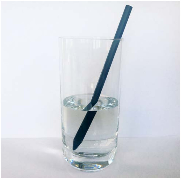
Fig. 1 A pencil image in two interfaces, air, and water.
With the use of a diving mask, a significant visual field reduction will also be present.5
With a standard diving mask, the horizontal visual field will go from about 180 ° in the air to about 97.2 ° underwater.3
The vertical field of vision will also decrease, with the reduction in the inferior field being the most likely to be of greater functional significance, since this may result in divers experiencing difficult in hand-eye coordination and consequently visualization of the equipment.17
The underwater perception of colors also changes, depending on the depth, due to the different wavelengths of visible light that are selectively absorbed, as visible light propagates in the water.17
Light rays with the highest wavelengths are absorbed first.
Red, for example, disappears at 9 meters and yellow at 23 meters.
At greater depths, these colors appear gray or black because the wavelengths responsible for these colors are absorbed by the water column.
At depths below 30 meters, there are only the blue and green components of visible light.6,17 (Figure 2)
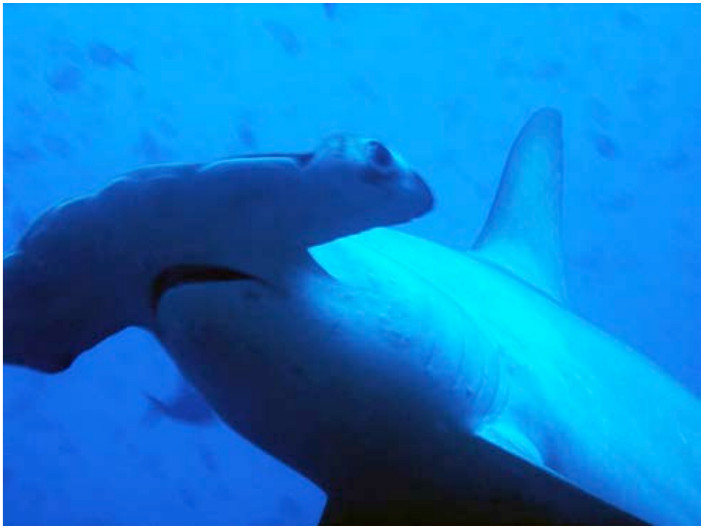
Fig 2 – Shark Hammer photographed below 30 meters.
The introduction of artificial light reverses this effect of color loss, at greater depths and in this way one can photograph the objects in their real color. (Figs 3 and 4)
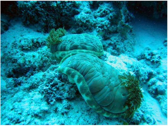
Fig. 3 - Nudibranch Hexabranchus sanguineus (Spanish Ballerina), without flash…
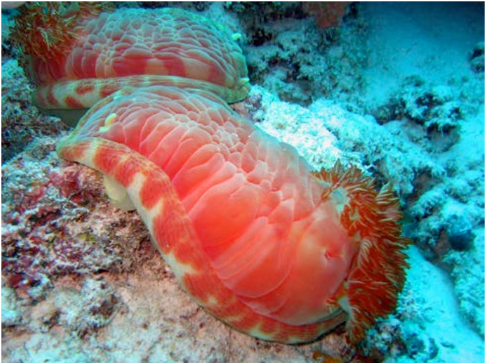
Fig. 4 – … and with flash!
Underwater Refractive Correction
If the diver has a refractive error, it is important to correct it not only to allow proper reading of the equipment (pressure gauge, depth gauge, watch, etc.) but also to enjoy the diving site.
There are several options for refractive correction:
1. Contact lenses: hydrophilic lenses are preferable since they allow the exchange of inert gases freely.7-11
Rigid gas permeable and rigid (PMMA) lenses may also be used if the dive is not too deep. Otherwise, corneal edema may appear during decompression and after diving.12,13
This occurs due to the development of nitrogen bubbles in the precorneal tear film, which interferes with normal tear physiology resulting in epithelial edema.14
The most frequent complication of contact lenses wear in diving is losing the lenses.15,16
This risk can be minimized by ensuring that the diving mask fits well. If water floods the mask while diving, half closing the eyes can also avoid losing the contact lenses.15
2. Replace the neutral lenses of the diving mask with corrected lenses, with the desired refractive prescription.
3. "Glue" spectacle lenses to the inner surface of the diving mask (although erosion or blistering may occur in the substance used to glue the lenses). (Fig 5)
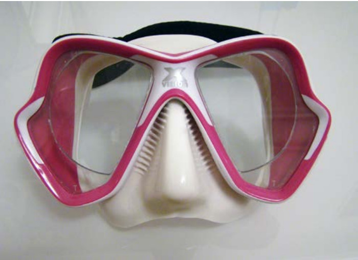
Fig. 5 - Diving mask with corrected spectacle lenses on the inner surface.
Diving after eye surgery
It is possible to dive after most eye surgeries.
However, the time required to wound healing must be guaranteed, before resuming diving. The waiting time is variable between different surgical procedures (but for most procedures, it is reasonable to wait between 3 to 6 months).7,18
In cases of glaucoma filtering surgery, in which a communication is created between the anterior chamber and the subconjunctival space, a barotrauma caused by the mask may have a negative effect on the bleb filtration mechanism and may lead to an additional surgical procedure or a possible exacerbation of the glaucomatous lesion.18
“One Sunday morning in 1936 at Le Mourillon, near Toulun, I waded into the Mediterranean and looked into it through Fernez goggles.
Merely a salty obstacle that burned my eyes.
I was astounded by what I saw in the shallow shingle at Le Mourillon, rocks covered with green, brown and silver forests of algae and fishes unknown to me, swimming in crystalline water.
Standing up to breathe I saw a trolley car, people, electric- light poles.
I put my eyes under again and civilization vanished with one last bow.
I was in a jungle never seen by those who floated on the opaque roof."
Jacques Cousteau
The Silent World